DNA Methylation
DNA methylation is a covalent epigenetic modification involving the transfer of a methyl group (-CH3) onto a DNA base, primarily targeting the 5-carbon position of cytosine within cytosine-phosphate-guanine (CpG) dinucleotides to yield 5-methylcytosine (5mC). This mark alters local chromatin structure and steric accessibility to modulate gene expression without altering the primary nucleotide sequence.
1. Molecular Structure and Chemical Catalysis
In mammals, DNA methylation occurs predominantly on cytosine within symmetrical CpG dinucleotides:
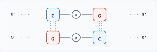
The modification is catalyzed by DNA methyltransferases (DNMTs) utilizing S-adenosylmethionine (SAM) as the universal methyl donor.
Catalytic Mechanism (C5-Cytosine Transfer)
- Base Flipping: The targeted cytosine is rotated out of the DNA double helix into the catalytic pocket of the enzyme.
- Nucleophilic Attack: A conserved cysteine thiolate in the active site executes a nucleophilic attack on carbon 6 (C6) of the pyrimidine ring.
- Enamine Intermediate: Formation of the covalent adduct at C6 shifts electron density to carbon 5 (C5), imparting nucleophilic character to C5.
- Methyl Transfer: Activated C5 attacks the electrophilic sulfonium methyl group of SAM, forming a covalent C-CH3 bond and generating S-adenosylhomocysteine (SAH).
- Beta-Elimination: Base-catalyzed abstraction of the hydrogen at C5 triggers beta-elimination, re-establishing the C5=C6 double bond and releasing the cysteine enzyme.
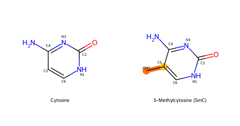
Because C5 projects outward into the major groove of B-form DNA, the addition of -CH3 does not disrupt Watson-Crick hydrogen bonding (O2, N3, and 4-NH2) between cytosine and guanine. Instead, it alters steric and electrostatic features recognized by DNA-binding proteins.
2. Enzymatic Machinery
Mammalian methylation involves distinct classes of enzymes responsible for establishment, maintenance, and removal.
A. De Novo Methyltransferases (DNMT3A and DNMT3B)
- Target unmethylated CpG dinucleotides to establish new methylation patterns.
- Active during early embryonic development, gametogenesis, and cellular differentiation.
- Guided by DNMT3L (a catalytically inactive regulatory cofactor) and specific unmethylated histone marks (such as unmethylated H3K4).
B. Maintenance Methyltransferase (DNMT1)
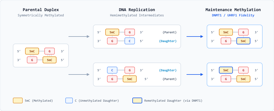
- Preserves established methylation patterns through mitotic cell divisions.
- Displays a 30- to 40-fold preference for hemimethylated CpG sites generated during semiconservative DNA replication.
- Recruited to replication forks via interaction with UHRF1 (Ubiquitin-like with PHD and RING finger domains 1), which recognizes hemimethylated DNA and H3K9me3.
C. Active Demethylation (The TET Family)
Methylation is reversed actively through iterative oxidation by Ten-Eleven Translocation (TET1, TET2, TET3) enzymes. These are Fe(II) and alpha-ketoglutarate-dependent dioxygenases that convert 5mC sequentially:
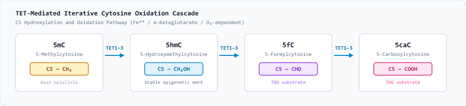
5fC and 5caC are recognized and excised by Thymine DNA Glycosylase (TDG). The resulting abasic site is repaired by the Base Excision Repair (BER) pathway, restoring an unmethylated cytosine.
3. Genomic Distribution and CpG Islands
Across mammalian genomes, CpG dinucleotides occur at roughly 20% to 25% of their statistically expected frequency. This genome-wide depletion is driven by differential susceptibility to spontaneous hydrolytic deamination:
- Unmethylated Cytosine \(\to\) Uracil (U): Hydrolytic deamination converts unmethylated cytosine into uracil, generating a foreign U:G mispair. Because uracil is non-canonical in DNA, it is promptly recognized and excised with high fidelity by Uracil-DNA Glycosylase (UDG) via the base excision repair (BER) pathway, restoring the original C:G pair.
- 5-Methylcytosine (5mC) \(\to\) Thymine (T): Deamination of 5mC at the C5 position yields thymine, generating a natural T:G base mismatch rather than an aberrant nucleobase. Repair by mismatch-specific glycosylases (such as TDG) is substantially less efficient, resulting in persistent evolutionary \(\text{C} \to \text{T}\) (and complementary \(\text{G} \to \text{A}\)) transition mutations over evolutionary timescales.
CpG Islands: Architecture and Regulatory Roles
Despite widespread genomic depletion, high CpG densities are preserved within defined, non-random clusters termed CpG Islands (CGIs).
| Parameter | Standard Definitional Criteria & Characteristics |
|---|---|
| Length | Typically 500 to 2,000 base pairs (minimum 200 bp). |
| Sequence Composition | \(\text{GC content} \ge 50\%\); ratio of observed-to-expected CpGs (\(\text{Obs/Exp}\)) \(> 0.60\). |
| Genomic Distribution | Intersect the 5′ transcription start sites (TSS) and promoters of approximately 70% of annotated human protein-coding genes. |
| Basal Epigenetic State | Maintained predominantly unmethylated across developmental stages and somatic tissues—including poised or transcriptionally silent tissue-specific promoters—shielded by open chromatin architecture and CxxC domain-containing factors (e.g., KDM2A, CFP1). |
| Repressive State | Aberrant or developmental de novo hypermethylation recruits methyl-CpG-binding domain proteins (MBDs, MeCP2) and histone deacetylase (HDAC) corepressor complexes, enforcing dense chromatin compaction and irreversible transcriptional silencing (e.g., genomic imprinting, X-chromosome inactivation, and pathological silencing of tumor suppressor genes in neoplasia). |
4. Mechanisms of Methylation-Mediated Transcriptional Silencing
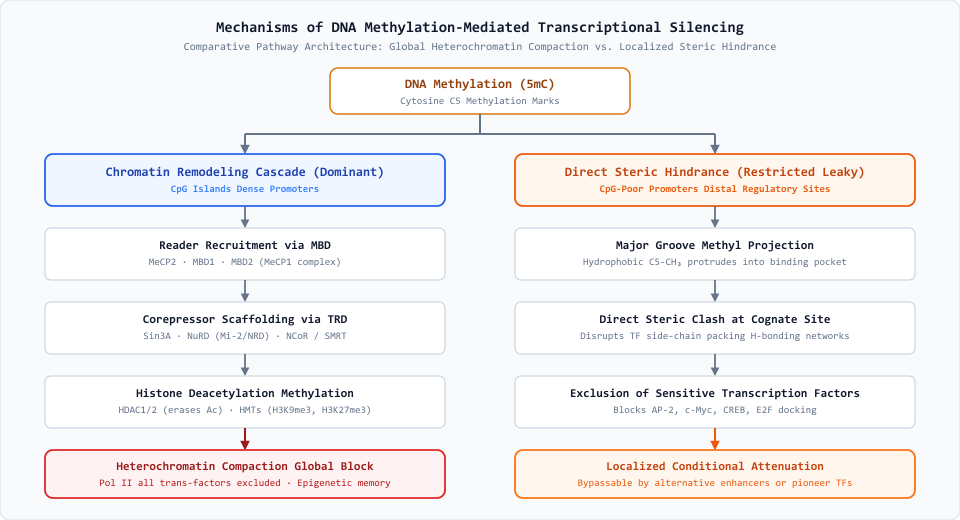
DNA methylation induces gene silencing through two distinct functional pathways: an indirect chromatin remodeling cascade and direct steric obstruction. While both modulate transcription, the chromatin remodeling pathway exerts far more robust, durable, and genome-wide repression.
The Dominant Mechanism: MBD Recruitment and Heterochromatin Assembly
The primary and most potent silencing route operates through cooperative assembly of repressive reader complexes across densely methylated CpG islands (CGIs). This occurs via a coordinated biochemical cascade:
- Epigenetic Tag Recognition: Methyl-CpG-binding domain (MBD) family members (including MeCP2, MBD1, and the MBD2-containing MeCP1 complex) selectively recognize symmetrically methylated CpGs via their modular DNA-binding domain.
- Corepressor Docking: Bound MBD readers utilize their Transcriptional Repression Domain (TRD) as a physical scaffold to recruit multisubunit corepressor complexes, predominantly Sin3A, NuRD (Mi-2/NRD), and NCoR/SMRT.
- Chromatin Modification:
* Deacetylation: Complex-associated Histone Deacetylases (HDAC1 and HDAC2) strip acetyl groups from histone amino-terminal tails (erasing activating marks like H3K9ac and H3K27ac), increasing the electrostatic affinity between basic histone octamers and the acidic DNA backbone.
* Repressive Methylation: Co-recruited Histone Methyltransferases (HMTs, such as SUV39H1, G9a, and PRC2 components) deposit repressive lysine marks (notably H3K9me3 and H3K27me3). - Steric Lockdown: The locus condenses into tightly coiled heterochromatin. This macromolecular compaction physically excludes RNA Polymerase II and basal transcription initiation factors from the promoter, establishing permanent epigenetic silencing (as observed in genomic imprinting, X-chromosome inactivation, and stable tumor suppressor silencing).
The Alternative Mechanism: Direct Steric Hindrance
The secondary pathway acts through localized, physical interference with sequence-specific transcription factor (TF) docking, independent of chromatin remodeling:
- Major Groove Projection: The hydrophobic methyl group added to the C5 position of cytosine projects directly into the major groove of the DNA double helix.
- Binding Motif Disruption: When a transcription factor’s cognate motif contains an internal CpG dinucleotide (e.g., binding sites for AP-2, c-Myc, CREB, or E2F), the protruding methyl moiety clashes with amino acid side chains in the factor’s DNA-binding domain or alters critical hydrogen-bonding networks.
- Scope and Vulnerability: This mechanism operates mainly at CpG-poor or intermediate-density promoters that rely on single, isolated transcription factor binding events for basal transcription.
Comparative Efficacy: Why Chromatin Remodeling Dominates
The chromatin remodeling cascade provides far more complete and durable transcriptional shutdown than steric hindrance alone:
| Dimension | Chromatin Remodeling Pathway | Direct Steric Hindrance Pathway |
|---|---|---|
| Primary Genomic Domain | CpG Islands and high-density promoters | CpG-poor promoters and distal elements |
| Effector Machinery | MBDs, MeCP2, Sin3A, NuRD, HDAC1/2, HMTs | Direct spatial obstruction in the major groove |
| Repressive Scope | Global steric exclusion: nucleosome condensation blocks all trans-acting factors and Pol II | Factor-specific: blocks only methylation-sensitive TFs |
| Mitotic Stability | Locked and self-reinforcing: maintains durable epigenetic memory across cell divisions | Conditional and dynamic: susceptible to TF concentration shifts or pioneer TF access |
| Leakiness | Minimal to non-existent across methylated islands | Leaky; nearby unmethylated motifs or alternative enhancers can still drive transcription |